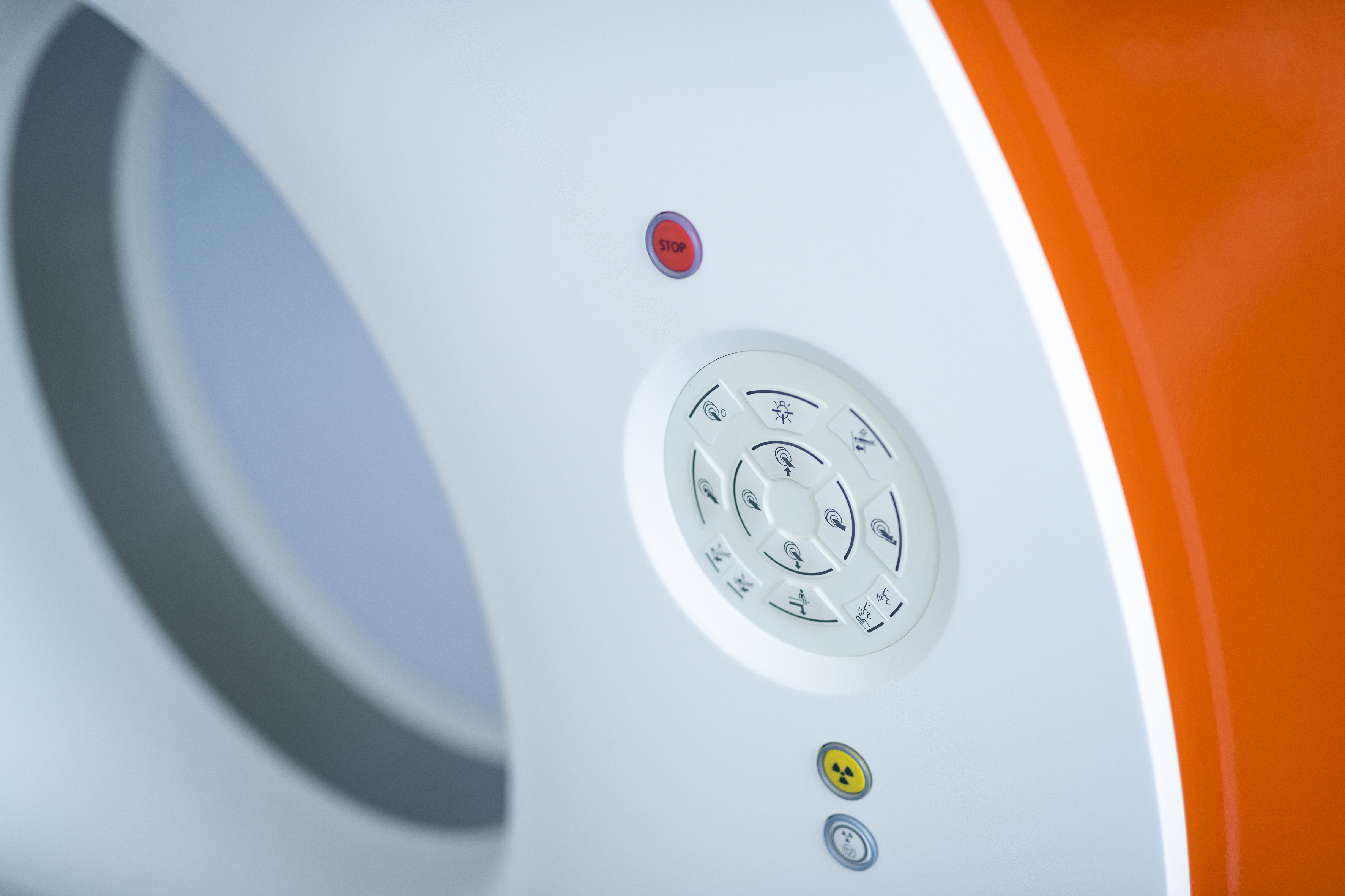
Computed tomography
Computed tomography
General information
The X-rays are measured as they pass through the body with detectors and then converted to cross-sectional images by computer (scan speeds of 2 to 350 milliseconds with simultaneous acquisition of up to 384 layers). The necessary radiation dose can be reduced by the latest technology by up to 90 percent.
The result: precise and overlay-free images that enable accurate evaluation and analysis; The smallest density differences – and thus the finest structures, such as in the lungs or in the abdomen – can be shown.
Special measuring programs allow the representation of the dental apparatus or the determination of the bone mineral salt content in osteoporosis examinations.
One of our highlights is our Dual Source Dual Energy CT at Diagnostikum Graz, a device equipped with two X-ray tubes. The result: double scanning speed, higher image quality, better presentation, more precise diagnostics despite up to 90 percent lower radiation dose.
- diagnosis of changes in the brain
- examination of diseases of the spinal column (e.g. intervertebral discs)
- diagnosis of diseases of the viscerocranium (e.g. paranasal sinuses) and the neck (e.g. larynx or lymph nodes)
- method to examine the fine structure of the lungs, the bones and the middle and inner ear
- evaluation of diseases of the thorax and the lungs
- examination of diseases of the abdominal organs (liver, pancreas, spleen, kidneys and adrenal glands)
- special examinations of the small intestine and the colon for treatment-resistant and unclear abdominal pain and chronic inflammatory bowel diseases
- the ideal method to image lymph nodes
- diagnosis of diseases of the pelvic organs (e.g. ovaries) and the pelvic soft tissues
- a safe method to image the large vessels (e.g. aorta or pulmonary arteries)
- dental CT: three-dimensional imaging of the upper and lower jaw and the teeth (e.g. before dental implants)
- osteoporosis CT: reliable and conclusive method to determine bone density
- cardio CT: the most reliable, non-invasive method to determine the calcium content of the coronary arteries and to rule out or provide evidence of a significant coronary heart disease
- virtual colonoscopy: four-dimensional imaging of the colon and the rectum to detect or rule out the presence of polyps and tumours (alternative diagnostic method to conventional endoscopy)
- low-dose multi-slice spiral CT of the lungs: currently the best method for early detection of bronchial carcinomas at the same exposure to radiation as in a thorax X-ray
- virtual angioscopy: three-dimensional imaging of the inner structure of the vessels
- virtual tracheobronchoscopy: four-dimensional imaging of the trachea and the bronchi to rule out or provide evidence of and for the surgical planning of bronchial tumours
- CT-targeted pain therapy: injection of medicines exact to the millimetre into the anatomical structures responsible for the pain, primarily the cervical spine, the lumbar spine and the sacroiliac joint
- CT-targeted organ biopsies: minimally invasive tissue extraction (e.g. from the lungs or the liver) for an exact histological characterisation