PET-CT
General Information
More details on PET-CT
Computed tomography is already a well-known diagnostic method in cross-sectional diagnostic imaging. It can be used to demonstrate the manifestation, morphology and location of a tumour in the body. Positron emission tomography (PET) provides information on the metabolic activity of the tumour, allows cancerous tissue to be found accurately in many cases and provides evidence of the effectiveness of chemotherapy. If the two techniques are combined, a unique method is created for precise tumour diagnostics and localisation, which supports the exact planning of individual therapy. PET-CT can also help in the decision of whether an operation or chemotherapy should be carried out.
Examination causes
- lung cancer
- breast cancer
- colorectal cancer
- melanoma
- malignant lymphoma
- head and neck cancer (incl. laryngeal cancer)
- oesophageal cancer
- pancreatic cancer
- thyroid cancer
- cervical caner, endometrial cancer (uterine cancer)
- ovarian cancer
- bladder cancer
- prostate cancer
Advantages of a PET-CT examination
- One single examination can be used to investigate whether metastasis of the tumour has already occurred.
- The effectiveness of chemotherapy can already be checked shortly after treatment has begun. If the glucose consumption of the tumour or metastasis has decreased, the cancer treatment is effective. However, if it has remained high, you can start using other treatment methods at an early stage and thereby improve your chance of recovery.
- During aftercare, it can be determined whether parts of the tumour or metastases have decreased or have recurred.
- You will receive a certain diagnosis in good time. This is important, since immediately beginning therapy in the treatment of cancer can often be decisive.
Equipment
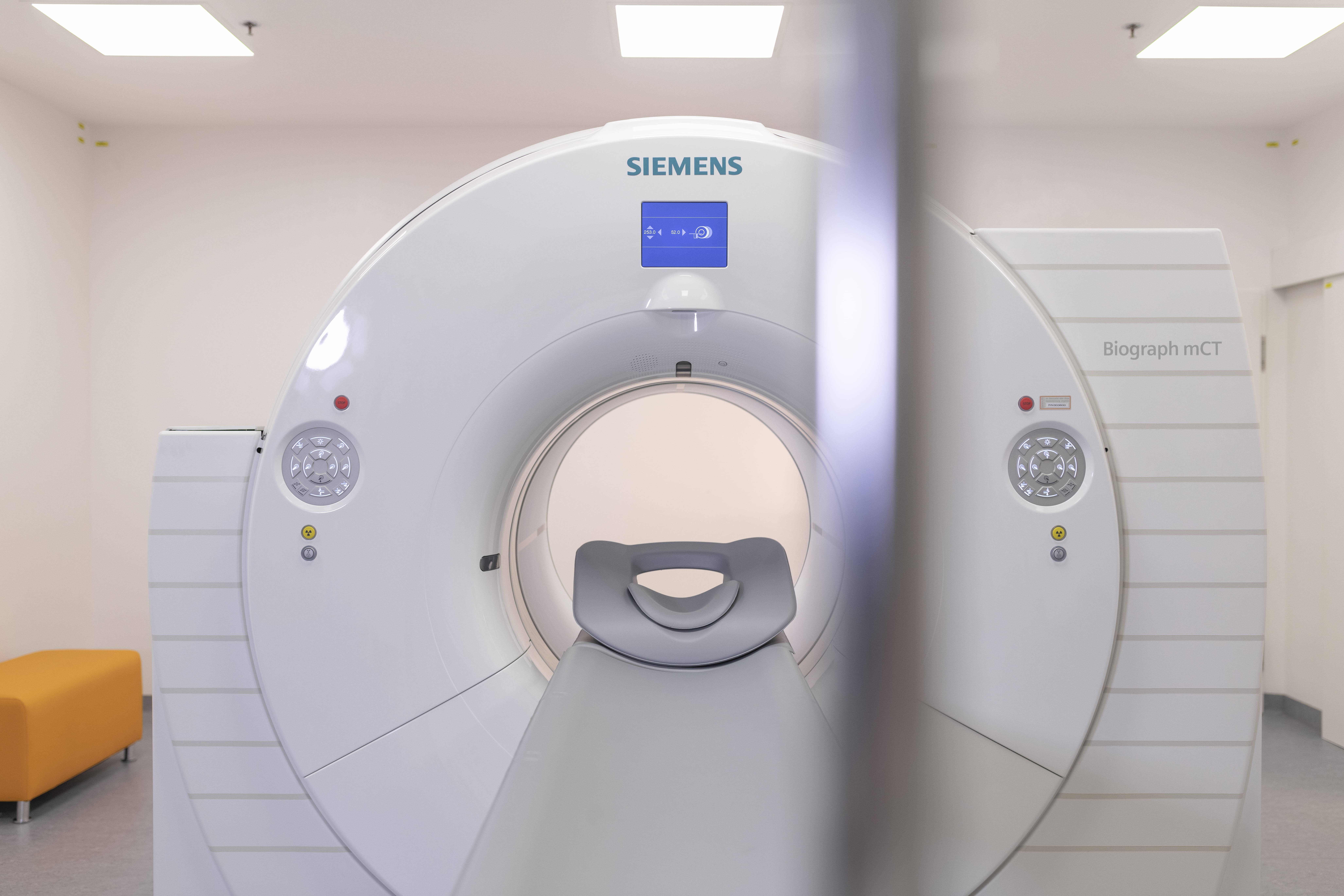
We operate one of the latest PET-CT scanners, which is equipped with innovative technology:
The highly sensitive detector allows for a reduction of the radiolabelled tracer dose with the shortest examination duration and the lowest radiation exposure.
The new calculation method ensures an exact measurement of the tumour’s activity.
The Siemens Biograph is one of our highlights at the institute in Graz.
You can find more information on PET-CT in Vienna (GE Discovery IQ), in cooperation with the diagnostic centres Urania and Favoriten at: petscan.at
How is the examination carried out?
For the PET-CT examination, you will be administered a weakly radioactive substance, a so-called tracer, into an arm vein. This is usually radiolabelled glucose, which makes metabolic processes in the body visible. It takes at least one hour until the tracer has accumulated in the body. We request that you lie still and relaxed during this time. During the examination itself, you lie on a sliding table that is moved through the PET-CT scanner.
How long does the examination take?
The examination itself usually takes no longer than around 30 minutes.
Is the examination painful?
Is the examination causing pain?
No, the examination, and particularly the administration of the tracer, is not painful. The tracer has no noticeable effect and is eliminated via the kidneys during the examination day.
